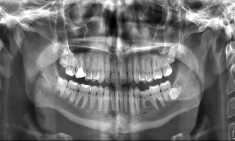
A dental x-ray is an essential tool to diagnose a diseased state of the oral cavity. A dentist can see the underlying structures like roots, any periapical lesion or bony status that are otherwise not visible in the oral cavity by doing an X-ray.
Dental X-rays
Nobody can oppose the significance of an x-ray for a bony fracture, but what about a dental X-ray? You will probably think why an x-ray is essential when a highly qualified dental surgeon is available for your intra-oral consultation.
The fact is that a dental x-ray is an essential tool to diagnose a diseased state of the oral cavity. By doing an X-Ray, a dentist can see the underlying structures like roots, any periapical lesion or bony status that are otherwise not visible in the oral cavity.
Dental x-rays are of two types:
- Intra-oral
- Extra-oral
Intraoral X-rays are preferred over the extra-oral ones in simple procedures that are limited to the oral cavity. Following are the three types of intraoral X rays:
- Periapical X-Ray view
- Occlusal x-ray view
- Bitewing x-ray view
Peri-apical X-ray
Also known as intraoral peri-apical (IOPA) view. This view can best view the areas that the tooth and its periapical area, regularity of lamina dura and periodontal ligamental space surrounding the alveolar bony status and root morphology.
This x-ray is most commonly used in dentistry, but the major drawback is that it can't be carried out in a limited mouth opening case.
Indications:
- To study the morphology of roots
- To check the angulation of impacted teeth
- Relationship of root with surrounding vital structures
- Dentoalveolar fracture
- To examine socket/ periapical lesion
- To explore the extent of caries
Occlusal X-ray view
Areas best seen in the maxilla are the hard palate and contour of the maxilla, while the areas that are best seen in the mandible are the floor of the mouth, the lingual aspect of the mandible, and its contour.
Indications:
- To study the number of ectopic or supernumerary teeth
- To study the impacted third molar
- Location of salivary calculus in submandibular gland's duct
- To examine the hard palate and its lesions
- To study the tumours arising on the lingual side of the tongue
When peri-apical and occlusal views are collectively considered, they will indicate the labio-lingual position of the tooth or the object.
Bitewing X-ray view
It is used to diagnose inter-dental caries, restoration overhangs, calculus deposits, and less than 4 mm bone loss. This radiographic view shows the crown of the tooth and the surrounding coastal bone level.

Dr Muntaha Tariq
The author is contributing writer at Dental News Pakistan and can be reached at muntahatariq@outlook.com